Sheep eyeball for home school – Welcome to the fascinating world of sheep eyeball dissection for homeschool! This engaging learning experience provides a hands-on exploration of the anatomy and function of the eye, offering valuable insights into the wonders of vision and the adaptations of living organisms.
As we delve into the intricate structure of the sheep eyeball, we’ll uncover the key components responsible for processing visual information and enabling sheep to navigate their environment. Through the process of dissection, we’ll gain a deeper understanding of the similarities and differences between sheep and human eyes, highlighting the adaptations that enhance vision in different species.
Introduction to Sheep Eyeball Anatomy

The sheep eyeball, a marvel of nature, is a complex organ responsible for vision and perception in these woolly creatures. Understanding its intricate structure and function is crucial for comprehending how sheep perceive their surroundings.The eyeball consists of several key components, each playing a vital role in the process of vision.
The cornea, a transparent outer layer, acts as a protective covering and allows light to enter the eye. Behind the cornea lies the lens, a flexible structure that adjusts its shape to focus light onto the retina. The pupil, a dark opening in the center of the iris, controls the amount of light entering the eye.
The iris, the colored part of the eye, regulates pupil size and contributes to eye color.At the back of the eyeball lies the retina, a delicate layer containing millions of light-sensitive cells called photoreceptors. These cells convert light into electrical signals that are transmitted to the brain via the optic nerve.
The brain interprets these signals, creating a visual representation of the world.Together, these components work harmoniously to enable sheep to see their surroundings, navigate their environment, and interact with their world. Understanding the anatomy of the sheep eyeball provides valuable insights into the sensory experiences of these fascinating animals.
Cornea, Sheep eyeball for home school
The cornea, the outermost layer of the eyeball, serves as a transparent protective shield. Its smooth, curved surface allows light to enter the eye and reach the inner structures. The cornea also helps focus incoming light, contributing to the eye’s ability to form clear images.
Lens
The lens, located behind the cornea, is a flexible, transparent structure that plays a crucial role in focusing light onto the retina. It changes its shape to adjust the focal length, ensuring that images are sharp and clear at varying distances.
Pupil
The pupil, a dark circular opening in the center of the iris, regulates the amount of light entering the eye. It contracts in bright light to reduce the amount of light reaching the retina and expands in dim light to increase light intake.
Iris
The iris, the colored part of the eye, surrounds the pupil and contains muscles that control its size. By adjusting the size of the pupil, the iris helps regulate the amount of light entering the eye and contributes to the eye’s ability to adapt to different light conditions.
Retina
The retina, lining the back of the eyeball, is a thin, light-sensitive layer that contains millions of photoreceptors. These cells, known as rods and cones, convert light into electrical signals that are transmitted to the brain via the optic nerve.
The brain interprets these signals, creating a visual representation of the world.
Dissecting a Sheep Eyeball

Dissecting a sheep eyeball is a valuable experience for students learning about the anatomy and function of the eye. By carefully following the steps Artikeld below, you can safely and effectively dissect a sheep eyeball to observe its internal structures.
Removing the Eyeball from the Socket
Before dissecting the eyeball, it must be carefully removed from the socket. Use sharp scissors to cut around the eyeball, taking care not to damage the optic nerve. Once the eyeball is free, gently pull it out of the socket.
Identifying External Features
Once the eyeball has been removed, take some time to identify its external features. The cornea is the clear, dome-shaped structure at the front of the eye. The iris is the colored part of the eye, and the pupil is the black hole in the center of the iris.
The sclera is the white, fibrous outer layer of the eye.
Separating and Examining the Eyeball Components
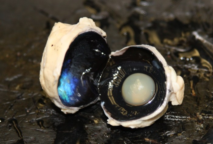
To dissect the eyeball, use a sharp scalpel to carefully cut through the sclera. Be careful not to cut into the lens or retina. Once the sclera has been cut, you can gently separate the different components of the eyeball.The
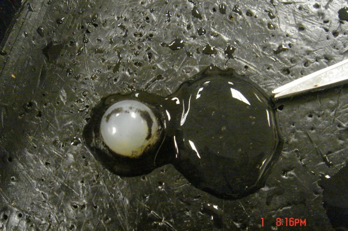
lens is a clear, biconvex structure that helps to focus light on the retina. The retina is a thin layer of tissue at the back of the eye that contains light-sensitive cells. The optic nerve is a bundle of nerve fibers that carries visual information from the retina to the brain.By
carefully dissecting a sheep eyeball, you can gain a better understanding of the anatomy and function of the eye. This experience can be a valuable addition to any science curriculum.
Comparing Sheep and Human Eyeballs

The eyes of sheep and humans share many similarities in their anatomy and function, enabling them to perform essential visual tasks. However, there are also some key differences between the two species’ eyeballs.
Dissecting a sheep eyeball is a fascinating home school science project that teaches kids about anatomy and biology. The experience is akin to reading chapter 4 of The Grapes of Wrath , where Steinbeck vividly describes the struggles of migrant workers.
Both endeavors provide an immersive and unforgettable learning journey that expands our understanding of the world.
The following table compares the anatomy and function of sheep and human eyeballs:
| Feature | Sheep Eyeball | Human Eyeball |
|---|---|---|
| Cornea | Transparent, outermost layer that covers the front of the eye, protecting the inner structures and focusing light onto the lens. | Similar to sheep’s cornea, it is responsible for focusing light onto the lens. |
| Lens | Biconvex, transparent structure behind the cornea that further focuses light onto the retina. | Similar to sheep’s lens, it adjusts its shape to fine-tune the focus of light onto the retina. |
| Pupil | Dark, central opening in the iris that allows light to enter the eye. | Similar to sheep’s pupil, it controls the amount of light entering the eye by adjusting its size. |
| Iris | Colored, muscular ring surrounding the pupil that controls the size of the pupil. | Similar to sheep’s iris, it contains muscles that contract and relax to change the pupil’s size. |
| Retina | Light-sensitive layer at the back of the eye that contains photoreceptor cells (rods and cones) that convert light into electrical signals. | Similar to sheep’s retina, it contains a higher concentration of cones in the central area, providing sharper central vision. |
| Other Features | – Tapetum lucidum: A reflective layer behind the retina that enhances night vision.
Sclera The tough, white outer layer of the eye that provides structural support. Optic nerve The bundle of nerve fibers that carries visual information from the retina to the brain. |
– Similar to sheep’s tapetum lucidum, it enhances night vision.
|
Overall, sheep and human eyeballs have similar structures and perform comparable functions, allowing both species to perceive and interpret visual information from their surroundings.
The Importance of Vision in Sheep

Vision is a crucial sense for sheep, enabling them to survive and thrive in their environment. Their eyesight plays a vital role in various aspects of their behavior, from navigating their surroundings to finding food and avoiding predators.
Sheep have evolved specific adaptations in their eyes that enhance their vision in different light conditions. These adaptations include:
Horizontal Pupils
- Sheep have horizontally elongated pupils, which provide them with a wider field of view. This allows them to scan their surroundings for potential threats or food sources while keeping their heads down to graze.
- The horizontal orientation of the pupils also reduces the amount of glare entering the eyes, improving their ability to see in bright sunlight.
Tapetum Lucidum
- The tapetum lucidum is a reflective layer located behind the retina in sheep eyes. It helps to reflect light back into the retina, increasing the amount of light available for vision.
- This adaptation enhances sheep’s night vision, allowing them to see in low-light conditions.
Using Sheep Eyeballs for Educational Purposes: Sheep Eyeball For Home School
Incorporating sheep eyeballs into homeschool curricula offers a unique and engaging approach to teaching principles of vision and anatomy. These specimens provide hands-on experiences that enhance understanding and foster scientific curiosity.
Sheep eyeballs are readily available and affordable, making them a practical choice for homeschool settings. They are comparable to human eyeballs in terms of structure and function, enabling students to make direct observations and draw meaningful connections.
Activities for Demonstrating Principles of Vision
1. Visual Acuity: Students can compare the sharpness of vision between the sheep and human eye by placing an object at varying distances and observing the clarity of the image.
2. Accommodation: By using a magnifying glass or microscope, students can demonstrate how the lens changes shape to focus on objects at different distances.
3. Field of View: Using two sheep eyeballs mounted on a model head, students can explore the binocular vision and wide field of view of sheep.
Activities for Exploring Anatomy
1. External Examination: Students can identify the major external structures of the eyeball, including the cornea, pupil, iris, and optic nerve.
2. Internal Dissection: With proper guidance, students can carefully dissect the eyeball to reveal the lens, retina, and vitreous humor.
3. Histological Examination: Advanced students can use a microscope to examine prepared slides of sheep eyeball tissues, observing the cellular components and their arrangement.
Benefits of Hands-on Learning
Hands-on experiences with real specimens, such as sheep eyeballs, offer several benefits for homeschool learners:
- Enhanced Understanding:Practical activities reinforce concepts and provide a deeper understanding of anatomical structures and their functions.
- Scientific Inquiry:Students engage in scientific inquiry by formulating hypotheses, conducting experiments, and drawing conclusions.
- Cognitive Development:Hands-on learning stimulates critical thinking, problem-solving, and spatial reasoning skills.
- Appreciation for Nature:Examining real specimens fosters an appreciation for the complexity and beauty of living organisms.
FAQ Corner
What safety precautions should be taken when dissecting a sheep eyeball?
Always wear gloves and safety glasses. Handle the eyeball with care to avoid puncturing or tearing. Dispose of the eyeball and dissection materials properly.
What are the benefits of using sheep eyeballs for homeschool science?
Sheep eyeballs provide a hands-on learning experience that allows students to explore the anatomy and function of the eye in a real-world context. They can observe the different components of the eye, learn about their roles in vision, and compare the structure of sheep eyes to human eyes.